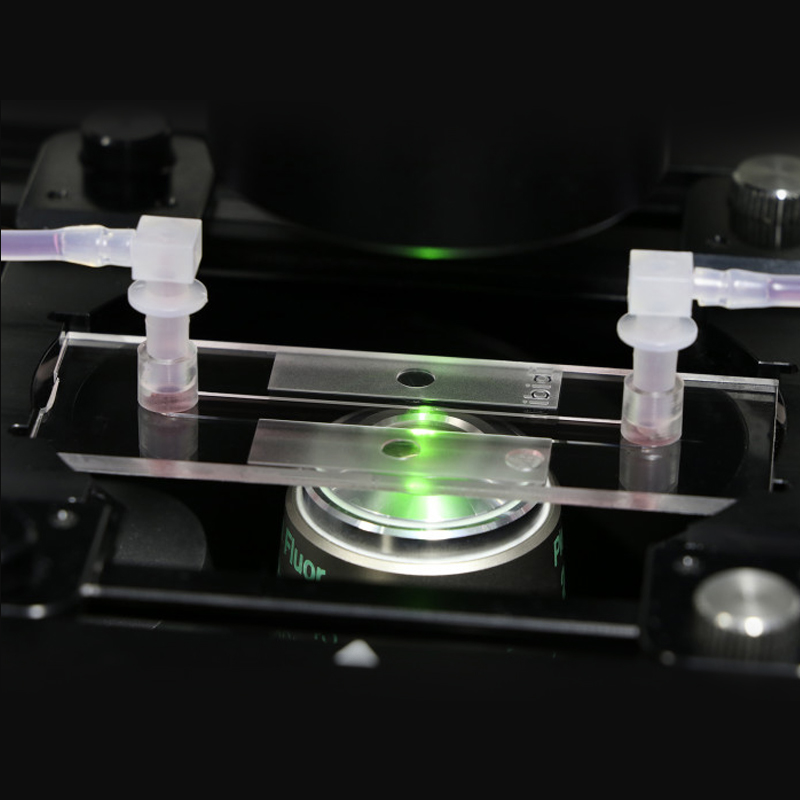


Adherent cells under flow conditions
Cell culture (static or stop-flow)
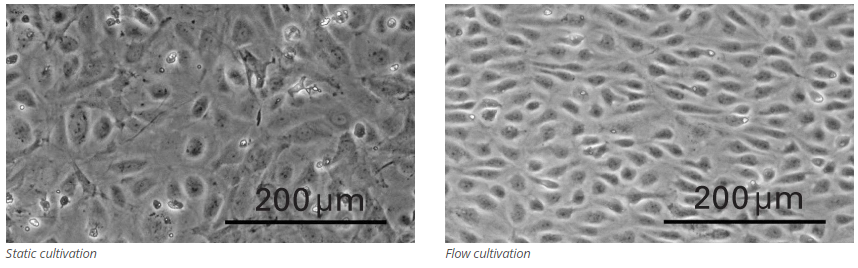
Simulation of blood vessels with endothelial cells
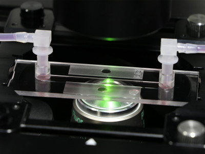
High-resolution microscopy of living and fixed cells
TIRF and super-resolution microscopy
Want to know if you should use a glass or a polymer bottom for your application? Find out here.
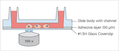
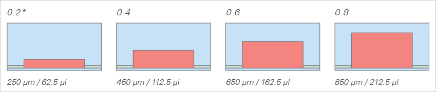
Because of the adhesive layer, the channel height of the μ-Slide I Luer Glass Bottom increases by 50 μm compared to the μ-Slide I Luer.
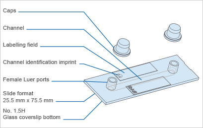
Specifications
Outer dimensions | 25.5 x 75.5 mm2 (w x l) |
Channel length | 50 mm |
Channel width | 5 mm |
Adapters | Female Luer |
Volume per reservoir | 60 μl |
Growth area | 2.5 cm2 |
Bottom: Glass coverslip No. 1.5H, selected quality, 170 μm +/- 5 μm | |
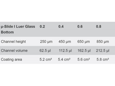
μ-Slide I Luer Glass Bottom | 0.2 | 0.4 | 0.6 | 0.8 |
Channel height | 250 μm | 450 μm | 650 μm | 850 μm |
Channel volume | 62.5 μl | 112.5 μl | 162.5 μl | 212.5 μl |
Coating area | 5.2 cm2 | 5.4 cm2 | 5.6 cm2 | 5.8 cm2 |
*Standard format with thin coverslip bottom made from D 263 M Schott glass #1.5H (170 μm +/- 5 μm) for low or high magnification microscopy
*Large observation area for microscopy
*Defined shear stress and shear rate levels
*Easy connection to tubes and pumps using Luer adapters
*Fully compatible with the ibidi Pump System
*May require coating to promote cell attachment
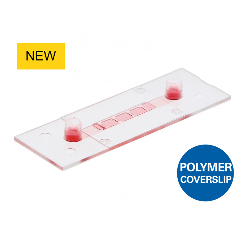
*Also available as a μ-Slide I Luer with an ibidi Polymer Coverslip Bottom for superior cell growth
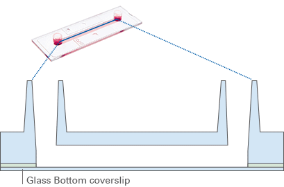
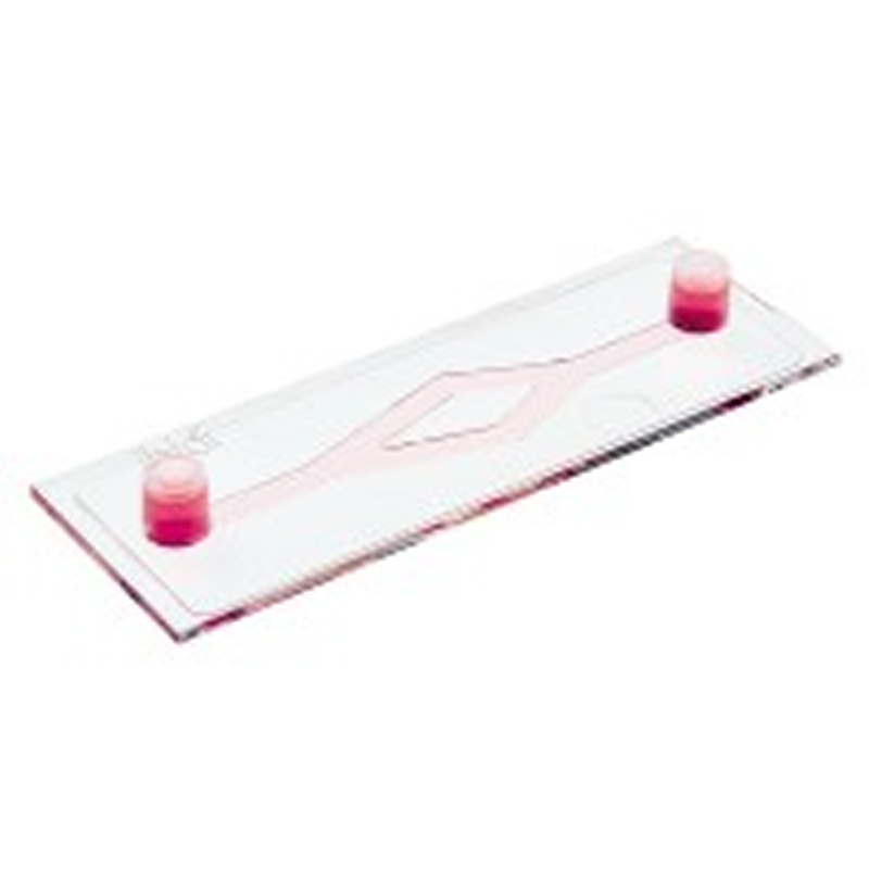
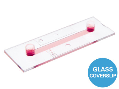
The Principle of the μ-Slide I Luer Glass Bottom
The Glass Bottom
The μ-Slide I Luer Glass Bottom comes with a thin #1.5H Glass Coverslip Bottom that has the highest optical quality and is ideally suitable for high-resolution microscopy, TIRF, and super-resolution microscopy. To promote cell attachment, a surface coating might be required prior to cell seeding.
Find more information and technical details about the coverslip bottom of the ibidi chambers here.